How Is Keratoconus Treated?
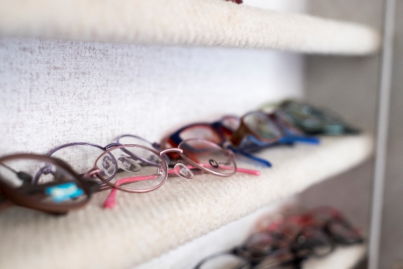
Spectacles/Eyeglasses
In the early stage and in mild keratoconus, eyeglasses may be all that is needed to improve vision. However, as the cornea becomes increasingly irregular, eyeglasses are less effective at correcting vision.

Contact Lenses:
Contact lenses (CL) work by creating an artificial, smooth surface on the front of the eye, improving the cornea’s ability to bend light. The majority of individuals with KC are prescribed CLs after their condition is diagnosed and continue to wear them successfully throughout their lives, allowing them to carry on normal productive lives.
Great care and expertise must be used by doctors who prescribe CLs for their KC patients. Frequent progress visits and changes in contact lens shape and power may be necessary due to changes in cornea shape. Specialty contact lens designs have been developed specifically for those with KC. These custom lenses may offer the best vision and comfort as KC progresses:
Soft Contact Lenses: Traditional disposable or soft contacts are not a typical option for KC patients, but some individuals with mild disease find these useful. There are also soft lenses made especially for keratoconus patients. They are useful for custom soft contacts may provide vision to those who find it difficult to tolerate ‘hard’ lenses. These lenses provide significantly less visual clarity, so the compromise between comfort and optimum vision must be carefully weighed.
Rigid Gas Permeable Lenses: The lens type most frequently used to correct KC are ‘rigid gas permeable’ or ‘gas permeable’ (RGP or GP) lenses. They provide excellent eye health because the lenses allow the cornea to ’breathe’ oxygen through the lens material. RGP lenses can be custom designed for the unique shape of the KC cornea and are easy to apply, remove, and care for. These lenses provide good vision correction, but some patients are unable to tolerate their wear over long periods of time.
Piggyback Lenses:A tandem or piggyback lens is a technique in which a soft contact lens is placed on the cornea and a corneal GP lens or hybrid lens sits on top of the soft lens. Although it takes more work to wear two lenses in oe eye, , some find this dual lens system prevents the rigid lens surface from irritating the sensitive cornea with the protective soft lens.
Hybrid Lenses: These specialty lenses incorporate a GP lens in the center, with a soft peripheral ‘skirt’. The hybrid lens offers the comfort, centration and stability of soft lens but the clear vision afforded by rigid central optics.
Scleral Lenses: Scleral lenses are large-diameter GP lenses, the size of a nickel to quarter, designed to vault over the entire cornea and rest on the sclera (the white part of the eye). Because of the size, the lens bowl must be filled with non-preserved saline before being placed on the eye. Individuals may initially find applying and removing scleral lenses challenging, but the majority achieve exceptional vision and comfort.
Intrastromal Corneal Ring Segments
Intrastromal corneal ring segments (ICRS) are clear, arc-shaped implants made of synthetic material that are surgically placed into precise tunnels created in the outer edge of the cornea by laser or other surgical instrument. ICRS implantation helps to remodel very steep corneas, making them more symmetric and flattening the curvature. In the U.S., these rings are called Intacs®. ICRS do not stop progression of KC. So, the goals of ring segments and corneal crosslinking are distinct. ICRS are designed to improve the corneal optics and reduce refractive error. Crosslinking has the goal of decreasing progression of disease. Some doctors combine ICRS placement and crosslinking into a single treatment; however no significant studies have been published to date with results for this combined procedure. Small scale trials have not demonstrated both procedures performed together to be superior. Even after insertion of ring segments, patients should expect to wear glasses or contacts for vision correction.
Not every patient with KC is a candidate for Intacs®. If this is a treatment option you would like to consider, please ask your eye doctor.
Corneal Crosslinking
Corneal Crosslinking (CXL) represents an important milestone in the treatment of keratoconus. While CXL has been performed for more than two decades internationally, approval by the U.S. Food and Drug Administration took place in 2016.
CXL is a nonsurgical procedure performed in the doctor’s office that takes about an hour. The treatment strengthens the weak corneal structure by allowing collagen fibers in the stroma to form new bonds to each other.
The result is that the progression of KC stops or is slowed. CXL does not reverse KC changes that have already occurred. That is why this procedure is recommended for those who are recently diagnosed or whose KC is still progressing. The procedure is less impactful for those who are no longer experiencing vision changes due to KC.
The only method that currently has FDA approval (as of the printing of this edition) utilizes an instrument manufactured by Avedro, Inc. (Waltham, MA) to deliver ultraviolet light and eye drops containing vitamin B2 (riboflavin). The treatment involves removing the central epithelium (the outermost layer of the cornea) to assure penetration of the eye drops.
This is called the ‘epi-off’ or epithelium-off method and is the standard CXL method. Eye surgeons are testing CXL protocols that do not require disturbing the epithelium (‘epi-on’). These treatments have yet not been shown to be as effective as the epi-off method and still have experimental or investigational status.
Following CXL, patients are told to expect a temporary decrease in vision and increased sensitivity to light for 1-3 months while the eye heals. Vision generally returns to pre-treatment levels in 6-12 months. New glasses and/or contact lenses are often required after treatment. The benefit of CXL is that further vision distortions slow or stop in the majority of cases.
Visit our Answering Your Questions about Crosslinking page for a list of common FAQ’s regarding CXL.
It has been more than 80 years since the first corneal transplant was performed on a patient with keratoconus. It remains the standard of care for the most severe cases, where comfort and useful vision cannot be achieved with other methods.
The tissue used for corneal transplants is donated from deceased organ donors. Corneas are removed within hours of death and are tested to assure they are disease-free and suitable for surgery. Transplants can last decades with proper care, but individual results vary.
The Penetrating Keratoplasty (PK or PKP) is the traditional transplant procedure and patients are likely to obtain excellent correctable vision. A trephine or surgical cookie-cutter is used to remove the entire full-thickness of the cornea (from epithelium to endothelium). The damaged cornea is replaced with similarly sized ‘button’ of donor tissue. Depending on the surgeon’s style, a series of individual sutures or one continuous suture holds the transplant in place. Over the next one-to-two years, the stitches are removed as the eye heals and vision improves. Patients are usually given prescription eye drops for an extended period of time to prevent rejection of the transplanted tissue.
In an increasing number of cases, the removal of the KC cornea and the preparation of the donor cornea are done using a femtosecond laser instead of a trephine. The procedure is referred to as FLEK (Femtosecond-Laser Enabled Keratoplasty). The laser is a precise cutting instrument, and can create more accurate, customized patterns in both the donor and recipient corneas. Laser assisted surgery may lead to faster wound healing and stronger bonds, resulting in lower astigmatism and better vision outcomes.
Another newer technique called Deep Anterior Lamellar Keratoplasty (DALK) has become a popular alternative among expert corneal surgeons. This is a more-delicate “partial-thickness” transplant, where the top or outer layers of the damaged cornea are replaced, but the Descemet’s membrane and endothelium are left in place. The DALK procedure is less invasive than a PK; there is a quicker recovery time and a decreased chance of graft rejection.
After corneal transplant surgery, whether full- or partial-thickness, the corneal surface irregularities may be reduced, but you will likely still need vision correction. With a new cornea, contact lenses are often better tolerated.
In a small number of cases, the transplant may fail or be rejected, and your surgeon may need to perform a repeat surgery. Infections are another potential complication you will need to be aware of. There have even been a few extremely rare reports of patients whose keratoconus recurs in the corneal transplant.
Many patients fear they will need to undergo a corneal transplant when they learn they have keratoconus. In most cases, there will not be a need for a transplant: more than 80% of individuals with KC do not require a corneal transplant. However, if needed, corneal transplantation using current eye-banking and surgical techniques is a very successful procedure.

